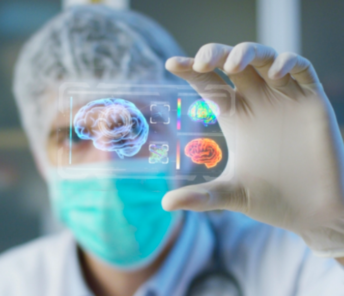
On the ever-changing horizon of developments in the imaging of brain injuries, one approach recently generated international interest. It was not the launch of another piece of sophisticated new imaging technology kit, but a simple blood test.
It was developed in the US working with injured veterans via the Department of Defence (DoD) and researchers from the Transforming Research and Clinical Knowledge in Traumatic Brain Injury (TRACK-TBI) Network using the principle of measuring biomarkers. The test measures two types of proteins, GFAP and UCH-L1, that are released from the brain and into the blood when the brain is injured.
A strength of the test, called the i-STAT® Alinity® system, is in its simplicity and utility: it is contained in a hand-held device, providing results within minutes. Trials are ongoing to see how the accuracy of the blood test system compares with results from CT scans; it is a promising but unproven addition to the brain injury assessment methods.
The assessment of brain injury is an area where the weaknesses of currently available imaging technology are widely recognised. In UK hospitals, if a scan is considered necessary when a patient presents to A&E, this will be a CT scan to help determine the extent of the injury, risk of complications and whether surgery may be required.
CT stands for computerised tomography (CT), using x-rays and a computer to create detailed images of the inside of the body. CT is the main imaging tool of hospital emergency departments; it is effective at identifying fractures and severe bleeds and is useful in the first 48 hours after an injury.
However, CT is less effective at showing damage within the brain where they may not be bleeding, such as injuries to microscopic nerve fibres. Research shows CT scans will miss a large number of mild Traumatic Brain Injuries (mTBI): an estimated 80 to 90 per cent of injuries will not be visible on standard CT imaging. Equally, the injury may produce damage within the brain which continues to take place after the initial time of presentation at an emergency department in hospital.
Here, MRI (magnetic resonance imaging) can be helpful and is often used to help explain enduring symptoms when the CT scan is clear. MRI is an area of rapid development, with variations in weight, diffusion and the enhancement of image quality improving the ability of scans to detect damage and changes at a microscopic level.
The range of technology introduced in recent years is broad, encompassing terms such as MR spectroscopy, Diffusion Weight imaging (DWI), Diffusion Tensor Imaging (DTI) / Diffusion Kurtosis Imaging (DKI), perfusion imaging, PET/SPECT, and magnetoencephalography (MEG).
Of course, for the affected individual and their family, this is very challenging to understand and navigate. Patients are heavily dependent upon the NHS services available within their area and the imaging available to services where they receive care. But for some, it can be valuable to explore other imaging options and second opinions.
We can help, advise and signpost families to specialist MRI imaging which may not be available within their local service, but could play an essential role in establishing a full and precise diagnosis. Although there is still some progress to be made, imaging technology in brain injuries is rapidly developing and particularly for individuals living with difficult symptoms without an accurate diagnosis, imaging can be essential in developing a more effective, targeted treatment plan.
For our terms of use and disclaimer follow this link: https://coulthursts.co.uk/
